Scanning electron microscopes
Scanning electron microscopes for use
Device Profile:
1. HITACHI S-3000N, Japan
1.Accelerating voltage: 0.3 - 30 kV
2.Lamp silk: Tungsten wire
3.Resolving power: 3.0 nm
4.Magnification: 15 – 300,000
5.Image capture: CCD-Camera EDS (HORIBA EX-250)
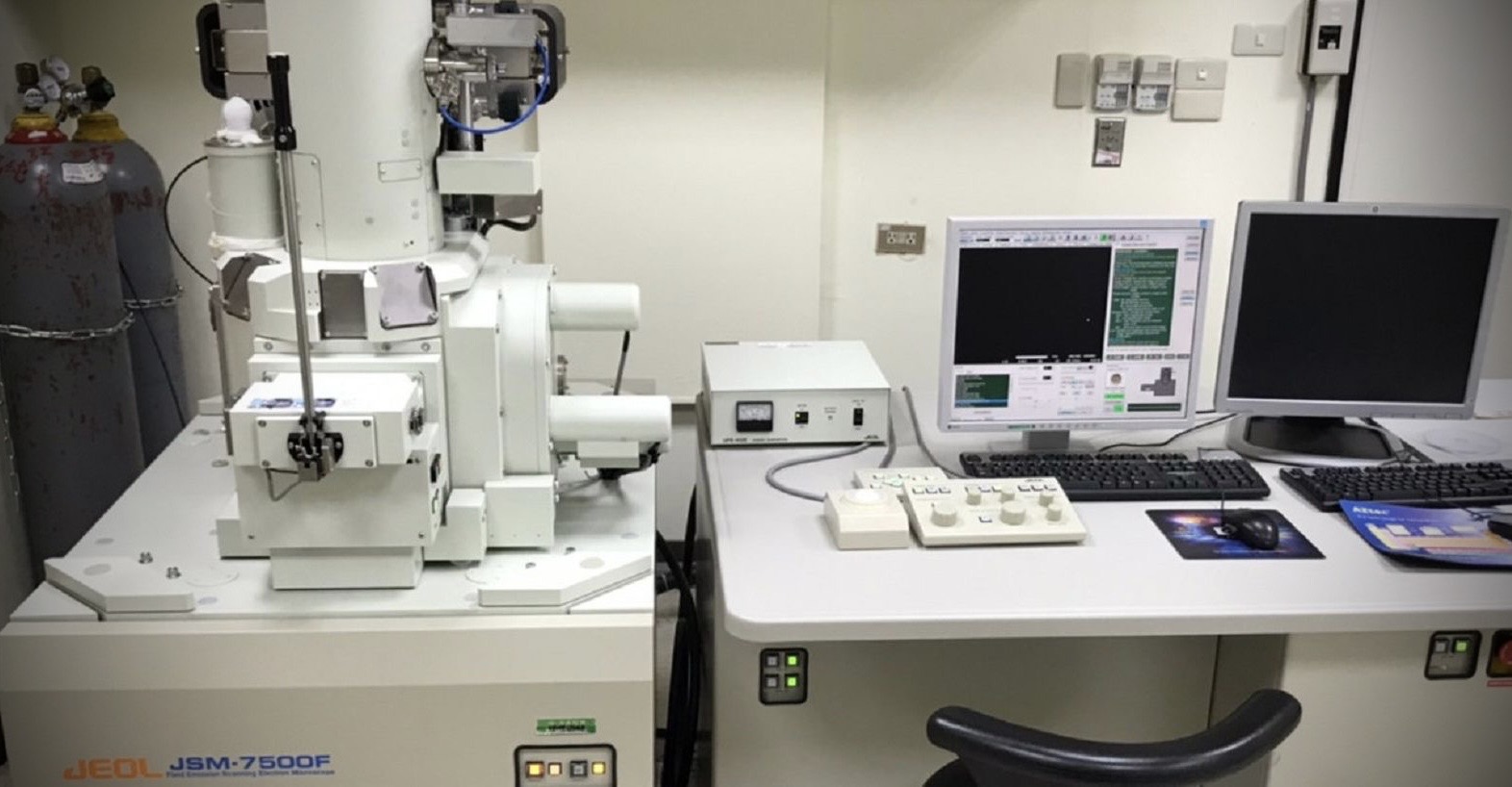
2. JEOL JSM-7500F, Japan
1.Accelerating voltage: 0.3 - 30 kV
2.Lamp silk: W(310) cold field emission
3.Resolving power: 1.0 nm
4.Magnification: 25 – 1,000,000
5.Image capture: CCD-Camera EDS (OXFORD INCA X-ACT)
Location:
The scanning electron microscopes room, Room 0459, 4thfloor of the 1st Medical Sciences Building.
Opening hours:
Monday to Friday, AM 8:30 ~ PM 5:00
Terms of Use:
Contact our technical staff to make an appointment for the use of the machine. Then fill out an application form. Arrive at the lab to use the machine at the scheduled time.
Charging method:
Fee list CGU Labs / Non CGU Labs
HITACHI S-3000N : 300 NT / 600 NT
JEOL JSM-7500F : 1200 NT / 2400 NT
Note: A period of less than half an hour counts as half-hour session. You will not be charged for any extended session of previous users
Notes:
1. We may reassign your pre-booked session to other users if you are late for more than one hour.
2. You will be disqualified from using the facilities for 6 months if you violate the terms and conditions for three times.
3. Experimental results provided by this center are for academic research only. The Center is not liable for any legal responsibility.
4. Fee and charges are subject to change. Please check our website for the latest fee information.
5. Power on and off: Please follow the correct procedure.
6. Data backup: (1)After each experiment, please immediately save experiment data. (2)The administrator clears the user data in the computer every six months.
Contact:
Mr. Peng
Tel: (03) 211-8800 ext 5065
E-mail: pon@mail.cgu.edu.tw
Device Profile:
1. HITACHI S-3000N, Japan
1.Accelerating voltage: 0.3 - 30 kV
2.Lamp silk: Tungsten wire
3.Resolving power: 3.0 nm
4.Magnification: 15 – 300,000
5.Image capture: CCD-Camera EDS (HORIBA EX-250)
2. JEOL JSM-7500F, Japan
1.Accelerating voltage: 0.3 - 30 kV
2.Lamp silk: W(310) cold field emission
3.Resolving power: 1.0 nm
4.Magnification: 25 – 1,000,000
5.Image capture: CCD-Camera EDS (OXFORD INCA X-ACT)
Location:
The scanning electron microscopes room, Room 0459, 4thfloor of the 1st Medical Sciences Building.
Opening hours:
Monday to Friday, AM 8:30 ~ PM 5:00
Terms of Use:
Contact our technical staff to make an appointment for the use of the machine. Then fill out an application form. Arrive at the lab to use the machine at the scheduled time.
Charging method:
Fee list CGU Labs / Non CGU Labs
HITACHI S-3000N : 300 NT / 600 NT
JEOL JSM-7500F : 1200 NT / 2400 NT
Note: A period of less than half an hour counts as half-hour session. You will not be charged for any extended session of previous users
Notes:
1. We may reassign your pre-booked session to other users if you are late for more than one hour.
2. You will be disqualified from using the facilities for 6 months if you violate the terms and conditions for three times.
3. Experimental results provided by this center are for academic research only. The Center is not liable for any legal responsibility.
4. Fee and charges are subject to change. Please check our website for the latest fee information.
5. Power on and off: Please follow the correct procedure.
6. Data backup: (1)After each experiment, please immediately save experiment data. (2)The administrator clears the user data in the computer every six months.
Contact:
Mr. Peng
Tel: (03) 211-8800 ext 5065
E-mail: pon@mail.cgu.edu.tw